
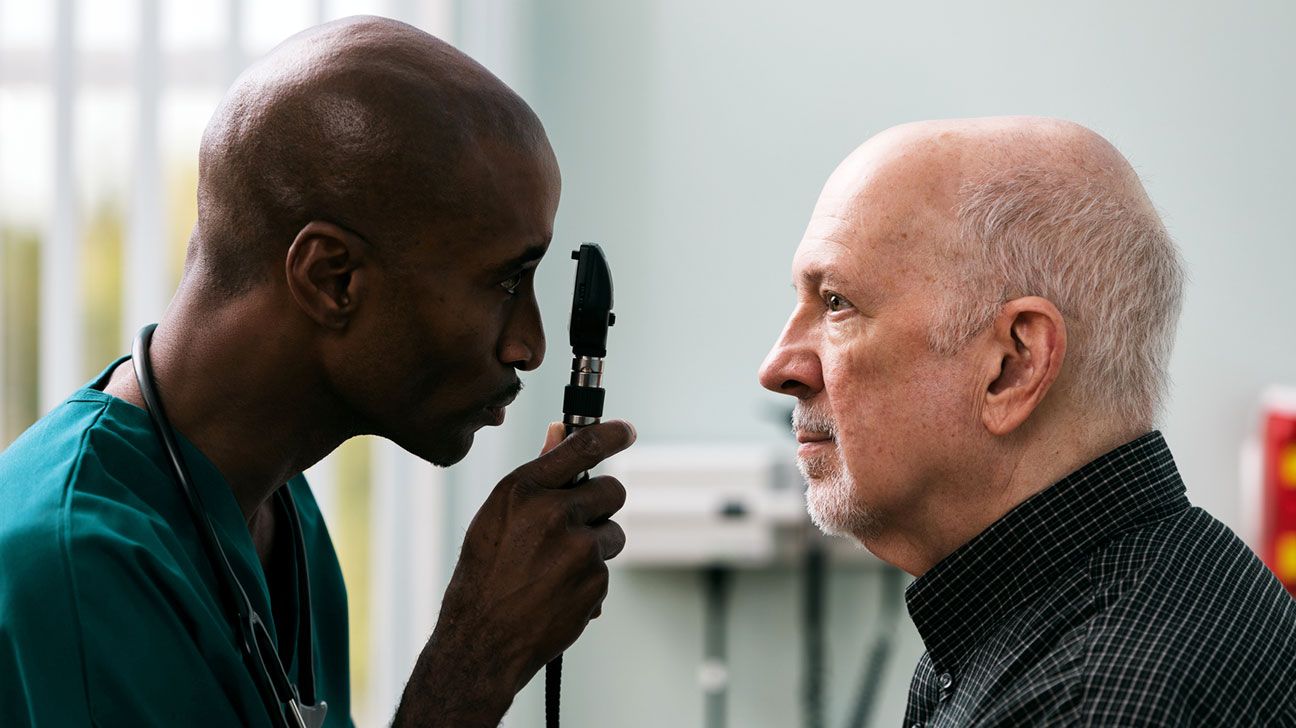
Fundoscopy can detect diabetic retinopathy. The exam involves a bright light shined into the eye, allowing an eye doctor to see any potential issues happening in the back of the eye.
Diabetic retinopathy is a common diabetes-related eye complication. To detect it in its earliest stages, eye doctors called ophthalmologists use an eye exam called fundoscopy.
Fundoscopy helps them diagnose and treat early retinopathy, slowing or stopping the progression of diabetic retinopathy and helping prevent vision loss.
This article explains what fundoscopy is and how it works.
Diabetic retinopathy is common in people with diabetes. Around
Fundoscopy, also known as ophthalmoscopy, is a medical test eye doctors use to diagnose retinopathy and other eye issues.
The exam can help eye doctors visualize the retina using an ophthalmoscope, which is no larger than a flashlight. It can diagnose health complications and risks of vision loss in the farthest reaches of the retina and surrounding areas, including the optic disk, choroid, and blood vessels.
There are three kinds of fundoscopies:
- Direct fundoscopy: While you sit in a dark room, your eye doctor shines a bright light through your pupil using an ophthalmoscope. Using this tool, they can see into the fundus of the eye to detect changes and complications.
- Indirect fundoscopy: Your eye doctor opens your eye and shines a bright light into your pupil using an ophthalmoscope strapped to their head. They will look into the fundus of your eye with the light by using a lens held close to your eye. Your eye doctor may also apply pressure to the eye using a small probing device. This type of fundoscopy can detect detached retinas.
- Slit-lamp fundoscopy: For this type, you sit in a chair and lean into an instrument in front of you. You rest your chin and head onto a support to keep your eye steady. Your eye doctor then uses a slit-lamp microscope and a lens in front of your eye to look into the retina. This type of fundoscopy provides greater magnification than an indirect fundoscopy and can also detect a detached retina.
Fundoscopies are not painful. They’re typically done after the eyes are dilated. The test only takes 5–10 minutes, plus the time for the eye dilation drops to take effect, typically up to 30 minutes. You may be sensitive to light for an hour or two after eye dilation.
Doctors recommend people with diabetes get a complete retina examination every year.
Fundoscopy can diagnose retinopathy and other diseases such as:
- inflammation
- infection
- glaucoma
- high blood pressure in your eye
- ocular melanoma
- optic nerve problems
Fundoscopy can be used solo or with other diagnostic tools.
When used by a licensed eye doctor, fundoscopes and other eye exam tools can detect diabetes eye complications and retinopathy most of the time.
However, fundoscopies are extremely accurate.
It can detect diabetic retinopathy in its earliest stages, with the potential for timely diagnosis and treatment to stop the condition’s progression.
Furthermore, fundoscopies can detect other eye abnormalities in the asymptomatic stage, making earlier diagnosis and treatment possible.
If you have diabetes, a fundoscopy is a routine part of your annual eye exam.
Visiting your eye doctor regularly can help them monitor changes to your retina. Routine visits allow your doctor to help curb or stop the progression of the earliest stages of diabetic retinopathy, even before symptoms begin.
If your fundoscopy results are abnormal, you may also be experiencing the following symptoms of diabetic retinopathy:
- blurry vision
- fluctuating vision
- floating, dark spots in your vision
- dark or empty areas in your field of vision
- vision loss
Diabetic retinopathy is a common complication of diabetes. A retinopathy fundoscopy is a diagnostic exam that can monitor changes to the retina, the deepest part of your eye.
In people with diabetes, prolonged high blood sugar levels can damage this delicate area of blood vessels.
The exam typically takes 5–10 minutes, plus the time it takes for any eye dilation drops to take effect. The exam can help your eye doctor diagnose diabetic retinopathy before symptoms even develop.